For a PDF Version of this Article Please Click Here
How To Improve Your Histology Fixation Process
Histology can be a tricky process to get right. Between working with very thin and delicate structures, and getting the perfect amount to sample, there is plenty of room for error. That’s why our resident Boekel Histologist partner shared three fixation tips to ensure a quality sample, each time.
There are three major parts to chemical fixation:
- Temperature
- Timing
- Solution
What Are The Aspects of Chemical Fixation?
Temperature
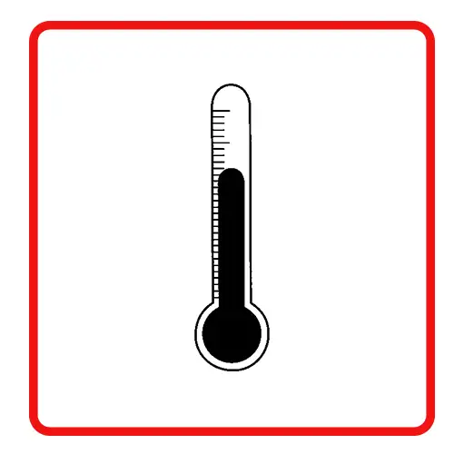
Maintaining an appropriate temperature during chemical fixation is crucial to the chemical reactions induced by the fixative. Room temperature, ranging from 20 to 25° C (68 to 77° F), is best to preserve the details of tissue morphology. Increased temperature increases the rate of fixation, but may also increase the rate of autolysis, destroying important areas of the tissue. By having control over the temperature of the environment, you’re able to ensure the sample you’re preserving is structurally sound.
Timing

Chemical fixation effectively stops the chemical processes within the cells contained in the sample. Without fixation, release of enzymes within cell organelles will lead to tissue degradation. Specimens should be placed immediately in a fixative solution, with larger specimens cut into smaller sections to allow rapid fixation. Most tissues should remain in the fixative solution for at least 12-24 hours, although some tissues have fewer artifacts with fixation of 48 hours to one week. By letting your sample remain in solution for the appropriate amount of time, you are allowing the chemical fixation process to maximize sample preservation.
Solution
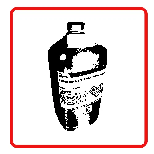
The correct solution should be used to process and stain the tissue sample for the intended evaluation. Tissue intended for immunohistochemical staining will require a different fixative than a sample intended for electron microscopy. A fixative solution contains a fixative like formalin dissolved in ethanol, water, or a buffer solution. A buffer solution contains a solvent such as water or alcohol combined with additional chemicals to maintain a constant pH through the fixation process. Some chemicals are included in a fixative solution to counteract the effects of other components. For example, because ethanol causes dehydration it will cause shrinkage to the tissue. This can be mitigated by adding acetic acid to the solution, as in Hartmann’s Fixative.
Carefully choosing the correct fixative for a process will lead to optimal results. Consult with histology guides and test different fixatives to determine the best solution for the process.
Contact Boekel Scientific
As you review your existing histology equipment and process, it might be time for an upgrade. Boekel Scientific offers a variety of equipment, reagents and consumables for histology. Contact us today or request a quote!